Opening up the natural world with new free to access digital images

How did the asteroid kill off the dinosaurs? By kicking up a huge amount of dust into the atmosphere, new research has suggested – Copyright GETTY IMAGES NORTH AMERICA/AFP Michael M. Santiago
Natural history specimens have never been so accessible following the completion of a project undertaken at the University of Texas at Arlington.
Biologists have painstakingly taken computed topography (CT) scans of more than 13,000 individual specimens. The digital images have then been used to to create 3D images of more than half of all the world’s animal groups, including mammals, fishes, amphibians and reptiles. This was made possible due to a grant from the U.S. National Science Foundation.
Computed tomography is a diagnostic imaging procedure that uses a combination of X-rays and computer technology to produce images of the inside of a body. It is possible to show detailed images, including the bones, muscles, fat, organs and blood vessels.
With the project, University of Texas worked with other institutions to develop the images. The images are now being populated, sone 30,000 media files in all, onto the open-source repository called MorphoSource.
This will allow researchers and scholars to share findings and improve access to material critical for scientific discovery.
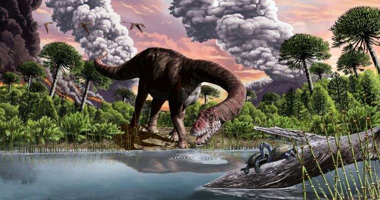
Among the findings of interest are using the data to conclude that Spinosaurus, a massive dinosaur that was larger than Tyrannosaurus rex and previously thought to be aquatic, would have actually been a poor swimmer, and thus likely stayed on land. Researchers have also revealed that frogs have evolved to gain and lose the ability to grow teeth more than any other animal.
Artists are also making use of the 3D models to create realistic animal replicas. Furthermore, photographs of the specimens have been displayed as part of museum exhibits. In addition, specimens have been incorporated into virtual reality headsets that allow users to interact with the animals. Educators also are able to use the digital models in their classrooms.
The project has been named the ‘openVertebrate project’ (or oVert). It means that anyone — scientists, researchers, students, teachers, artists — can look online to research the anatomy of just about any animal imaginable without leaving home.
In terms of specimen conservation, the digitalisation will also help curators to reduce wear and tear on many rare specimens while increasing access to them at the same time. The exercise in creating the images has been outlined in the journalBioScience, with the research paper titled “Increasing the impact of vertebrate scientific collections through 3D imaging: The openVertebrate (oVert) Thematic Collections Network.”
Opening up the natural world with new free to access digital images
#Opening #natural #world #free #access #digital #images


